The language of serotonin

Or, “What are they saying?

When we mention the word Serotonin (5-hydroxytryptamine, 5-HT), we immediately think of the brain and the Central Nervous System (CNS). People tend to associate serotonin to depression, or mood, or feelings of well-being1

Although that is correct, truth be told, the majority of the serotonin in the human body is actually produced in the gut. In fact, 95% of total serotonin is manufactured by the Enterochromaffin cells (or, Kulchitsky cells) in the gastro-intestinal tract (GI)2,3. These cells live next to the gut epithelium, that covers the cavity of the GI tract, playing a crucial role in the regulation of bowel movements and secretions. If you think that the gut is almost 9 meters (or 30 feet) long, then that’s a lot of cells producing serotonin. 

When in the 50’s, Betty M. Twarog and Irvine H. Page discovered that the brain produced its own serotonin4; then, the gut-made serotonin got reduced to its “Aschenputtel” origins, and relinquished to the favela quarters of the body. As such, brain-derived serotonin always got more attention than its gut-derived counterpart – like a rich vs. poor-cousin type of reputation.

Moving-on…

Platelets, also called thrombocytes, are small un-nucleated fragment of cells that, when activated, form blood clots (thrombus) and prevent bleeding. 

Electron microscopy images of circulating platelets, extracted from Zilla et al, 19875

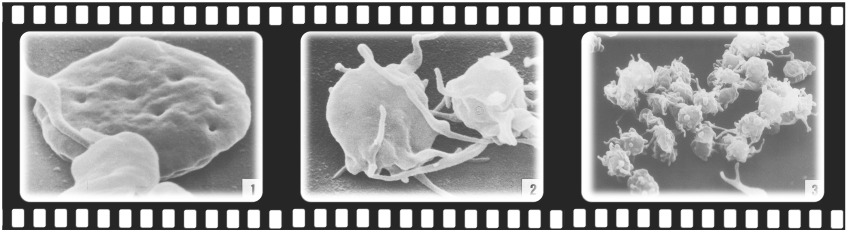
Platelets do not make serotonin, butcan take it up as they circulate through the gut, and carry it along the blood stream6,7. As such, the serotonin produced in the intestine can be carried all over the body. As the chemical messenger serotonin is, it can influence any other cell, in whatever other location, as long as it has a serotonin receptor on it. As such, peripheral serotonin has now discovered its path back into the limelight, and recent research has strengthened the influence that gut-made serotonin has in other parts of the body, functioning as an intestinal-derived hormone. 

Once again, the “Aschenputtel” story comes into mind, but this time through its “Cinderella” version. Let’s take a look…

For example, gut-derived serotonin can directly regulate the liver and mediate liver regeneration8. In Non-Alcoholic Fatty Liver Disease (NAFLD), a group of conditions that are characterized by excessive fat accumulation in the liver and closely track the global public health problem of obesity, researchers showed that inhibiting gut-derived serotonin synthesis could resolve hepatic fat accumulation8,9.

Peripheral serotonin can also be a negative regulator of bone density, by specifically inhibiting osteoblast formation and leading to osteoporosis10 – a common feature in patients with inflammatory bowel disease (IBD). This happens through the action of a common receptor: the low-density Lipoprotein Receptor-related Protein 5(LRP5), which is expressed in both osteoblasts and enterochromaffin cells11. LRP5 inhibits the expression of an important ingredient for serotonin production (Tryptophan hydroxylase-1, Tph1); as such, when LRP5 is deficient or inactivated due to inflammation or disease, blood levels of serotonin are elevated decreasing osteoblast formation; and, consequently, reducing bone mass1,11.

Epidemiologic data suggests a role of serotonin, or Selective Serotonin-Reuptake Inhibitors (typically used as antidepressants, SSRIs) in the development of venous thrombosis12. In fact, patients with depression were reported to have higher incidences of venous thromboembolism in general13; and, the use of SSRIs is associated with an increased venous thromboembolism risk14. No wonder, serotonin and platelets are “brothers in arms”, ready to block any blood vessel along their way…. 

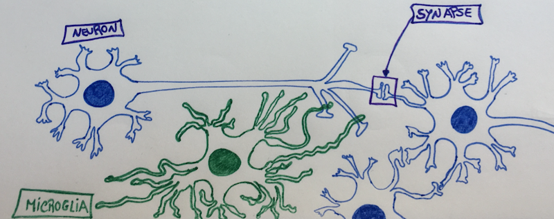
Serotonin and its receptors are also present in the immune system, where evidence suggests it contributes to both innate and adaptive responses. There is now clear evidence of a straight communication between the immune system, the gut and the brain via serotonin15,16.

On top of all and because we are not alone, our gut microbiota plays a critical role in regulating our colonic serotonin. Indigenous spore-forming bacteria (Sp) promote serotonin biosynthesis in our enterochromaffin cells, and with that they can significantly modulate GI movements and platelet function – together with many aspects of our physiology17,18. We now know that the microbiota colonizes the GI tract after birth, with a continuous maturation during the first years of life19. Researchers have now showed in animal models that this developing gut microbiota regulates the development of the adult enteric nervous system via intestinal serotonin networks20. What this actually means, is that the actions of our intestinal bugs during the beginning of our life are determinant for the development of our “gut brain”, our second brain. How about that?…

If we ruminate about it, when we “think” with our gut, we are actually listening to our bugs. By directly signalling our cells to produce serotonin and develop a network of neurons as soon as we are born, our gut-bugs are actually finding a way to communicate with us – the host – in the serotonin language. 

Now, we just need to understand what are they telling us… 

Beethoven’s hearing aids, Beethoven House Museum, Bonn.

References:

1          Gershon, M. D. 5-Hydroxytryptamine (serotonin) in the gastrointestinal tract. Curr Opin Endocrinol Diabetes Obes 20, 14-21, doi:10.1097/MED.0b013e32835bc703 (2013).

2          Bellono, N. W. et al. Enterochromaffin Cells Are Gut Chemosensors that Couple to Sensory Neural Pathways. Cell 170, 185-198.e116, doi:10.1016/j.cell.2017.05.034 (2017).

3          Yaghoubfar, R. et al. Modulation of serotonin signaling/metabolism by Akkermansia muciniphila and its extracellular vesicles through the gut-brain axis in mice. Scientific Reports 10, 22119, doi:10.1038/s41598-020-79171-8 (2020).

4          Twarog, B. M. & Page, I. H. Serotonin Content of Some Mammalian Tissues and Urine and a Method for Its Determination. American Journal of Physiology-Legacy Content 175, 157-161, doi:10.1152/ajplegacy.1953.175.1.157 (1953).

5          Zilla, P. et al. Scanning electron microscopy of circulating platelets reveals new aspects of platelet alteration during cardiopulmonary bypass operations. Tex Heart Inst J 14, 13-21 (1987).

6          Morrissey, J. J., Walker, M. N. & Lovenberg, W. The absence of tryptophan hydroxylase activity in blood platelets. Proc Soc Exp Biol Med 154, 496-499, doi:10.3181/00379727-154-39702 (1977).

7          Hughes, F. B. & Brodie, B. B. The mechanism of serotonin and catecholamine uptake by platelets. J Pharmacol Exp Ther 127, 96-102 (1959).

8          Wang, L. et al. Gut-Derived Serotonin Contributes to the Progression of Non-Alcoholic Steatohepatitis via the Liver HTR2A/PPARγ2 Pathway. Frontiers in Pharmacology 11, doi:10.3389/fphar.2020.00553 (2020).

9          Choi, W. et al. Serotonin signals through a gut-liver axis to regulate hepatic steatosis. Nature Communications 9, 4824, doi:10.1038/s41467-018-07287-7 (2018).

10        Lavoie, B. et al. Gut-derived serotonin contributes to bone deficits in colitis. Pharmacol Res 140, 75-84, doi:10.1016/j.phrs.2018.07.018 (2019).

11        Yadav, V. K. et al. Lrp5 controls bone formation by inhibiting serotonin synthesis in the duodenum. Cell 135, 825-837, doi:10.1016/j.cell.2008.09.059 (2008).

12        Rieder, M., Gauchel, N., Bode, C. & Duerschmied, D. Serotonin: a platelet hormone modulating cardiovascular disease. J Thromb Thrombolysis 52, 42-47, doi:10.1007/s11239-020-02331-0 (2021).

13        Takeshima, M. et al. Prevalence of Asymptomatic Venous Thromboembolism in Depressive Inpatients. Neuropsychiatr Dis Treat16, 579-587, doi:10.2147/NDT.S243308 (2020).

14        Parkin, L. et al. Antidepressants, Depression, and Venous Thromboembolism Risk: Large Prospective Study of UK Women. J Am Heart Assoc 6, doi:10.1161/jaha.116.005316 (2017).

15        Baganz, N. L. & Blakely, R. D. A dialogue between the immune system and brain, spoken in the language of serotonin. ACS Chem Neurosci 4, 48-63, doi:10.1021/cn300186b (2013).

16        Jacobson, A., Yang, D., Vella, M. & Chiu, I. M. The intestinal neuro-immune axis: crosstalk between neurons, immune cells, and microbes. Mucosal Immunology 14, 555-565, doi:10.1038/s41385-020-00368-1 (2021).

17        Yano, J. M. et al. Indigenous bacteria from the gut microbiota regulate host serotonin biosynthesis. Cell 161, 264-276, doi:10.1016/j.cell.2015.02.047 (2015).

18        Reigstad, C. S. et al. Gut microbes promote colonic serotonin production through an effect of short-chain fatty acids on enterochromaffin cells. Faseb j 29, 1395-1403, doi:10.1096/fj.14-259598 (2015).

19        Bäckhed, F. et al. Dynamics and Stabilization of the Human Gut Microbiome during the First Year of Life. Cell Host Microbe 17, 690-703, doi:10.1016/j.chom.2015.04.004 (2015).

20        De Vadder, F. et al. Gut microbiota regulates maturation of the adult enteric nervous system via enteric serotonin networks. Proc Natl Acad Sci U S A 115, 6458-6463, doi:10.1073/pnas.1720017115 (2018).

Skin microbiome: feed it right for a healthier look!

Dry skin and atopic dermatitis have been associated with changes in the variety of the skin microbiome. 

Our skin, as the largest organ in our body, has a huge array of commensal microbes that support a healthy skin barrier. One of those is Staphylococcus epidermidis, one of the most abundant bacterial species of the skin microbiome1.

This chubby mutualistic, Gram-positive, facultative anaerobe constitutes up to 90% of the aerobic resident flora of our skin, and has been associated with a healthy-looking skin2. It does not like to be lonely, and usually appears in pairs or tetrads on the surface of our skin, like a protecting biofilm.

Dry skin, for example, is associated with an increase in microbial diversity along with a decrease in microbial load in comparison to more sebaceous areas of the skin, that are usually populated by lipophilic bacteria such as Cutibacterium acnes – that tend to cause those unwanted teenager-look-a-like pimples that nobody likes…

Lactic Acid is one of the Natural Moisturizing Factors (NMF) of the skin barrier, that is essential to maintain the hydration and a slightly acidic pH of the skin surface (i.e., “acid mantle”)3. Higher lactic acid concentrations and lower skin surface pH are known to increase our epidermal renewal and promote a healthier skin. 

New in vitro data suggests that Staphylococcus epidermidis, may be one of the major sources of lactic acid in the skin1

But only if fed the right way. 

It seems that 1% colloidal oat increases Lactic Acid production by this particular bacteria species, making it rely less on simple sugars such as glucose for its metabolism; and, instead use more complex carbohydrates derived from oat.

Oatmeal-containing skin moisturisers significantly changed the metabolism of the Staphylococcus epidermidis, breaking down starch and promoting good gene expression, with an increased DNA and aminoacid synthesis, and an improved ATP metabolism.

How about that?

Bacteria on a diet makes your skin look healthier!

Next time you think about which moisturiser to buy in the drug store:  don’t forget to feed your skin microbiome it’s oatmeal!

Happy Staphys!

References:

1          Liu-Walsh, F. et al. Prebiotic Colloidal Oat Supports the Growth of Cutaneous Commensal Bacteria Including S. epidermidis and Enhances the Production of Lactic Acid. Clin Cosmet Investig Dermatol 14, 73-82, doi:10.2147/CCID.S253386 (2021).

2          Baviera, G. et al. Microbiota in healthy skin and in atopic eczema. Biomed Res Int 2014, 436921, doi:10.1155/2014/436921 (2014).

3          Thueson, D. O., Chan, E. K., Oechsli, L. M. & Hahn, G. S. The roles of pH and concentration in lactic acid-induced stimulation of epidermal turnover. Dermatol Surg 24, 641-645, doi:10.1111/j.1524-4725.1998.tb04221.x (1998).

Yogurt as precision medicine, or how your gut might be undermining your health

The gut microbiome is a community of microorganisms that lives in our gastrointestinal tract. It is so far, the most studied microbial community in healthy humans, because of its known role in a range of functions and diseases, like Inflammatory Bowel Disease (IBD)1,2.

To gain perspective on the magnitude of the bacterial presence inside of us, and potential effects on our bodies, the human body expresses 20,000 eukaryotic genes while the gut microbiome expresses 3.3 million prokaryotic genes. This suggests that the genetic contribution of the microbiome to humans may be many hundreds of times greater than the genetic contribution from the human genome.

Most of the microbes in the microbiome do not cause disease. In fact, we need them to perform many important functions that we cannot do ourselves. Microbes digest food to generate nutrients for host cells, synthesize vitamins, help to absorb nutrients and minerals, produce short-chain fatty acids, metabolize drugs, detoxify carcinogens, stimulate renewal of cells in the gut lining, and activate and support the immune system1

The fermentation by-products acetate, propionate, and butyrate are important for gut health; and, provide energy for epithelial cells, enhance the integrity of the epithelial barrier, and provide immunomodulation and protection against pathogens1

Current investigations explore resident bacterial gene function, and the potential role it might have in human health and metabolism. Each individual has its own microbiome, and no one common microbe is present in all body sites or all individuals. 

Researchers identified the composition of different individual microbiomes, but they also identified the metabolic pathways of the microbial communities found in different body sites (e.g., skin, colon, liver…).  What is interesting is that microbial membership diverges greatly between healthy individuals; but, the metabolic pathways of our own microbiomes is very similar, with common ‘housekeeping’ properties that maintain cell function and a functional body site ecosystem3,4.

The interactions between the gut microbiota and our bodies immune system begins at birth4. The microbiota shapes the development of the immune system; and, in turn, the immune system shapes the composition of the microbiota. This cross-talk between the microbes and our bodies is transmitted through a vast array of signaling pathways that involve many different classes of molecules, and extend upon multiple organs such as the gut, liver, muscle, and the brain. This creates axes of metabolic pathways, or highways of chemical communication, between the gut and the different organs in our bodies.

Because the gut microbiome is highly malleable, it can be altered throughout our lifespan by environmental factors, such as diet, stress and medication. What we have seen during the last 60 years, is an increaseincidence of gut dysbiosis, which is an imbalance in the intestinal bacteria that leads to disease.

As such, there is much interest in developing new therapeutic tools for manipulating the composition of the gut microbiota to benefit our health. A better understanding of how variations in this symbiotic relation within us, supraorganisms, will contribute to disease risk and health sustainability; and, will point the way to new therapeutic interventions and disease prevention strategies.

Danone, a leading yogurt multinational food corporation, is developing “precision probiotics”, for example. Researchers at Danone aim to tailor probiotics to an individual’s diet, phenotype, lifestyle, age, gender, genetics and microbiome. The intention it’s to bring to the gut activities or functions that are not provided by our own gut microbiome, or our own genes.

It’s funny that around 1920’s, Isaac Carasso, the creator of Danone, first started selling yogurt in pharmacies, using ferments isolated from the Institute Pasteur, and label it as health-food. It’s like going full circle.

References:

1          Bordigoni, A., Halary, S. & Desnues, C. in Encyclopedia of Virology (Fourth Edition) Vol. https://www.sciencedirect.com/topics/medicine-and-dentistry/gut-microbiome  (eds Dennis H. Bamford & Mark Zuckerman)  552-558 (Academic Press, 2021).

2          Lloyd-Price, J. et al. Multi-omics of the gut microbial ecosystem in inflammatory bowel diseases. Nature 569, 655-662, doi:10.1038/s41586-019-1237-9 (2019).

3          Visconti, A. et al. Interplay between the human gut microbiome and host metabolism. Nature Communications10, 4505, doi:10.1038/s41467-019-12476-z (2019).

4          Nicholson, J. K. et al. Host-Gut Microbiota Metabolic Interactions. Science 336, 1262-1267, doi:10.1126/science.1223813 (2012).

Feeling anxious or depressed? Might be your microglia…

A macrophage is a hungry immune cell that engulfs and eats all things that don’t have a good reputation in our body (e.g., cellular debris, pathogens…); and, microglia cells are the resident macrophage population of the Central Nervous System (CNS)1. They function as sentinels of local infection in the brain, backing both innate and adaptive immune responses, and account for 10-15% of all cells found in the brain and spinal cord2.

Microglia cells are also involved in the maintenance of brain homeostasis, contributing to mechanisms that underly learning and memory. They constantly survey their local microenvironment – like patrols – extending their motile processes, or hands/legs, to make a brief contact with neuronal synapses. This continuous synaptic plasticity, throughout our lifetime, is essential to control maladaptive learning and memory, such as addiction3. For example, the number of synapses in the brain regions of the nucleus accumbensamygdala and dorsomedial striatum increase when we expose our brains to addictive substances (such as alcohol, or opiates); and, decrease upon withdrawal due to the action of microglia cells4. As such, microglia cells help to modify and eliminate synaptic structures when they grow too much, or, are on the way to touch too many other neurons5 – because, neurons tend to be touchy and to enjoy a synaptic orgy. 

Whenever a neuron starts to freak out that it has too many synapses and it needs help regulating its neuronal “touchy” behaviour, then the synapse extends a greeting “hand” (filopodia) and “Hi5s” the neighbouring microglia cell, telling her that it needs help remodelling. Once “Hi5ed”, the microglia cell starts nibbling on the synapse6 – cutting all the excess – and, avoiding that that specific neuron gets assigned a bad “sexual” reputation. It’s like behaviour counselling, transforming and remodelling, but neuron-wise and with a microglia cell as the counsellor…

Even though microglia cells are essential and extremely helpful; like everything in life, they can also go haywire, ending up pruning too many synapses, and destroying healthy tissue. An uncontrolled activation of the microglia can be directly toxic to neurons, because they can release inflammatory cytokines (IL-1, TNF-alpha, IL-6, Nitric Oxide, Prostaglandine E2, and Superoxide)7, and lead to excessive pruning of neuronal synapses3.

The most recent research in the pathophysiology of depression and anxiety shows that abnormalities in microglia cells have a central role in the development of these diseases8. For example, a neuroimaging study in depressed patients, revealed that stronger depressive symptoms related with microglial activation in brain regions associated with mood regulation (the prefrontalanterior cingulate, and insular cortices of the brain)9. Additionally, post-mortem studies of depressed suicide victims showed microglial activation and macrophage accumulation within the anterior cingulate cortex brain region10

Persistent stress activates a chronic low-inflammatory state in our bodies that enhances our inflammatory response to challenges11. Social stress causes the release of inflammatory monocytes into the circulation8, which end up reaching the Blood Brain Barrier (BBB) and its endothelial cells. This low-systemic inflammation that travels through our vessels, encourages the migration of the brain resident microglia cells to the area of the cerebral vessels. In here, microglia cells make physical contact with endothelial cells of the BBB, and “sense” the inflammatory environment that is present in the blood (aka, inflammatory cytokines activate receptors in the microglia cells). If there is sustained inflammation, then some of the microglia cells can “become neurotic” and start nibbling the end-feet of healthy cells, making the BBB more permeable and, consequently, damaging the protective BBB shield function12. This is turn, leaks inflammatory cytokines from the blood into the brain tissue, further activating more microglia cells, that start cutting synapses from healthy neurons.

What this means is that a persistent low-grade inflammation can trigger microglia activation and change the functional connectivity of healthy neurons in major brain emotional centers13. Because our immune system can interact with the neurocircuitry that is involved in emotion regulation and behaviour, a chronic low-inflammation derived from stress can influence the development of various neuropsychiatric disorders, like depression and anxiety. 

But, what can we do to avoid falling in this trap?

Eat well, sleep well, do sports and have a good laugh with friends. All things that inhibit inflammation, and make us feel good. 

Microglia cell (green) “counselling” a synapse

References:

1.         Ginhoux F, Lim S, Hoeffel G, Low D, Huber T. Origin and differentiation of microglia. Frontiers in Cellular Neuroscience. 2013;7

2.         Lawson LJ, Perry VH, Gordon S. Turnover of resident microglia in the normal adult mouse brain. Neuroscience. 1992;48:405-415

3.         Neniskyte U, Gross CT. Errant gardeners: Glial-cell-dependent synaptic pruning and neurodevelopmental disorders. Nat Rev Neurosci. 2017;18:658-670

4.         Spiga S, Talani G, Mulas G, Licheri V, Fois GR, Muggironi G, et al. Hampered long-term depression and thin spine loss in the nucleus accumbens of ethanol-dependent rats. Proc Natl Acad Sci U S A. 2014;111:E3745-3754

5.         Tremblay M-È, Lowery RL, Majewska AK. Microglial interactions with synapses are modulated by visual experience. PLOS Biology. 2010;8:e1000527

6.         Weinhard L, di Bartolomei G, Bolasco G, Machado P, Schieber NL, Neniskyte U, et al. Microglia remodel synapses by presynaptic trogocytosis and spine head filopodia induction. Nature Communications. 2018;9:1228

7.         Kim YS, Joh TH. Microglia, major player in the brain inflammation: Their roles in the pathogenesis of parkinson’s disease. Exp Mol Med. 2006;38:333-347

8.         McKim DB, Weber MD, Niraula A, Sawicki CM, Liu X, Jarrett BL, et al. Microglial recruitment of il-1β-producing monocytes to brain endothelium causes stress-induced anxiety. Mol Psychiatry. 2018;23:1421-1431

9.         Setiawan E, Wilson AA, Mizrahi R, Rusjan PM, Miler L, Rajkowska G, et al. Role of translocator protein density, a marker of neuroinflammation, in the brain during major depressive episodes. JAMA Psychiatry. 2015;72:268-275

10.       Suzuki H, Ohgidani M, Kuwano N, Chrétien F, Lorin de la Grandmaison G, Onaya M, et al. Suicide and microglia: Recent findings and future perspectives based on human studies. Frontiers in cellular neuroscience. 2019;13:31-31

11.       Miller GE, Rohleder N, Cole SW. Chronic interpersonal stress predicts activation of pro- and anti-inflammatory signaling pathways 6 months later. Psychosom Med. 2009;71:57-62

12.       Haruwaka K, Ikegami A, Tachibana Y, Ohno N, Konishi H, Hashimoto A, et al. Dual microglia effects on blood brain barrier permeability induced by systemic inflammation. Nature communications. 2019;10:5816-5816

13.       Kim J, Yoon S, Lee S, Hong H, Ha E, Joo Y, et al. A double-hit of stress and low-grade inflammation on functional brain network mediates posttraumatic stress symptoms. Nature Communications. 2020;11:1898